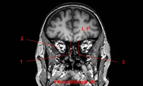
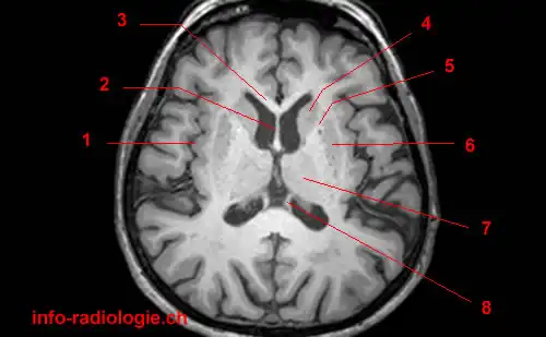
44 brain mri with labels
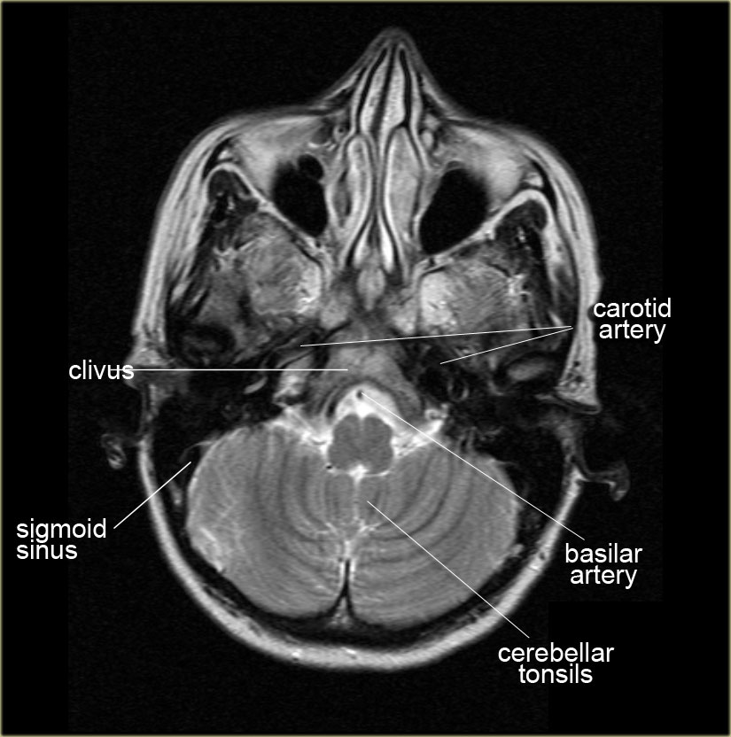
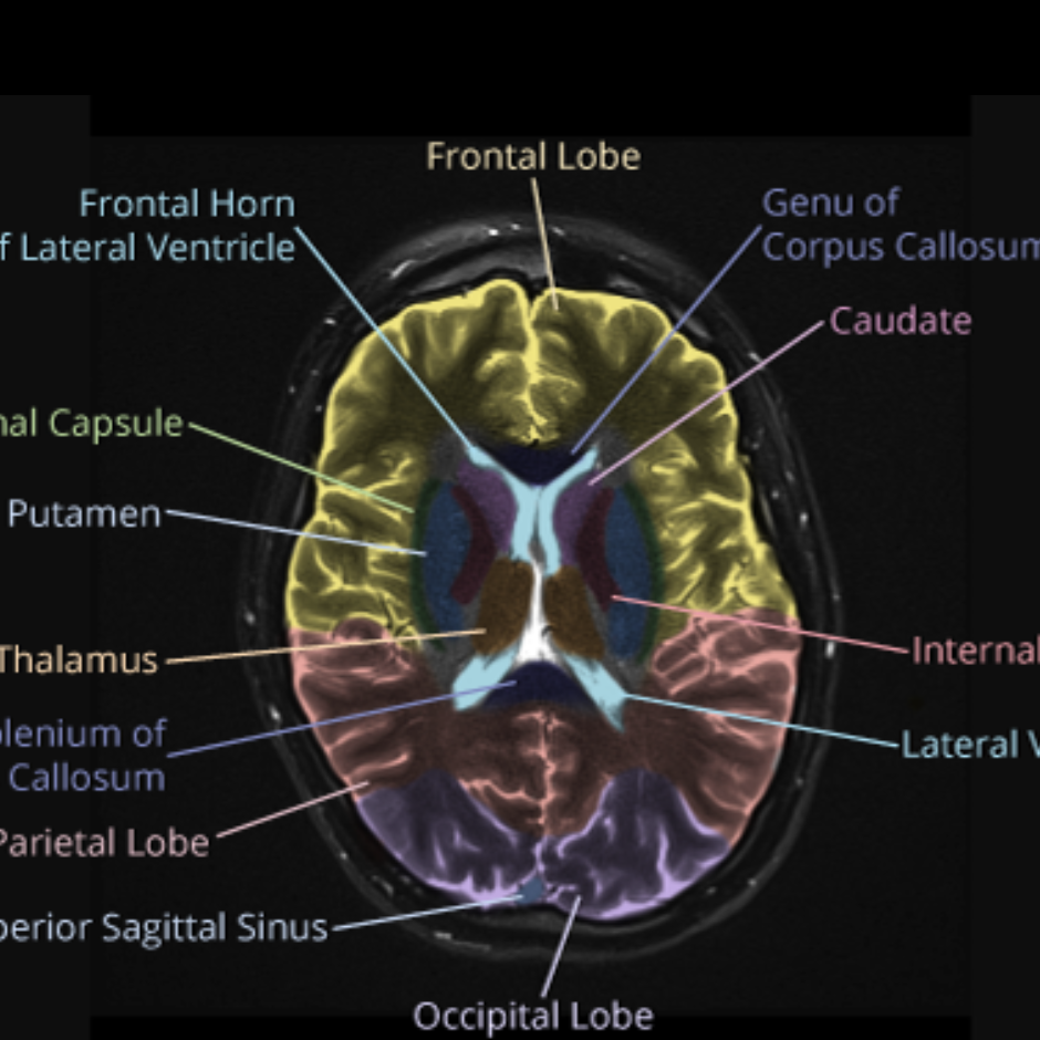
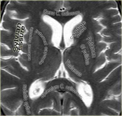
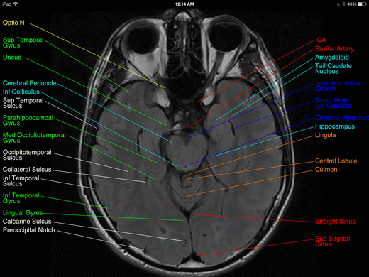
CaseStacks.com - MRI Brain Anatomy Labeled scrollable brain MRI covering anatomy with a level of detail appropriate for medical students. Show/Hide Labels. MRI Brain Anatomy. Back to Anatomy Overview. ... Labelled radiographs and CT/MRI series teaching anatomy with a level of detail appropriate for medical students and junior residents. Pelvis. Pelvic MRI anatomy › environmentEnvironment - The Telegraph Oct 22, 2022 · Water meters should be compulsory and bills should rise, says new Environment Agency chairman. Alan Lovell says households consume too much water and metering is needed to encourage them to cut ...
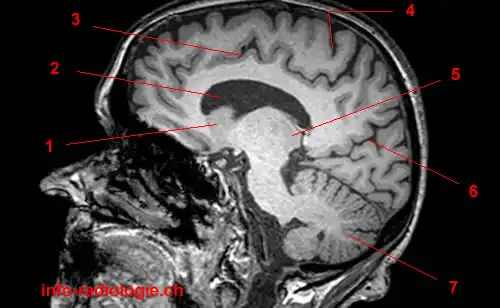
MRI head sagittal T1 - labeling questions | Radiology Case ... The labeled structures are (excluding the correct side): temporal horn of lateral ventricle primary fissure of cerebellum choroid plexus trigone (atrium) of lateral ventricle horizontal fissure of cerebellum occipital horn of lateral ventricle intraorbital segment of optic nerve diploic space of parietal bone body of caudate nucleus maxillary sinus

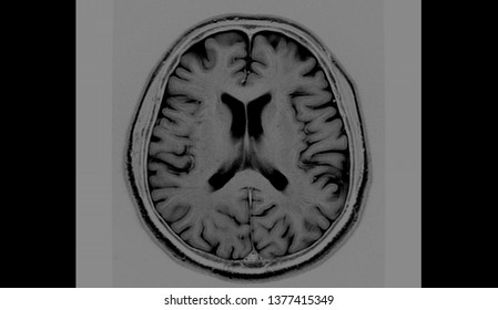
Brain mri with labels
Labeled imaging anatomy cases | Radiology Reference Article ... This article lists a series of labeled imaging anatomy cases by body region and modality. Brain CT head: non-contrast axial CT head: non-contrast coronal CT head: non-contrast sagittal CT head: angiogram axial CT head: angiogram coronal CT... Atlas of BRAIN MRI - W-Radiology Brain magnetic resonance imaging (MRI) is a common medical imaging method that allows clinicians to examine the brain's anatomy (1). It uses a magnetic field and radio waves to produce detailed images of the brain and the brainstem to detect various conditions (2). 101 labeled brain images and a consistent human cortical ... - PubMed We introduce the Mindboggle-101 dataset, the largest and most complete set of free, publicly accessible, manually labeled human brain images. To manually label the macroscopic anatomy in magnetic resonance images of 101 healthy participants, we created a new cortical labeling protocol that relies on robust anatomical landmarks and minimal manual edits after initialization with automated labels.
Brain mri with labels. Brain lobes - annotated MRI | Radiology Case | Radiopaedia.org Anatomy - Neuro, Head & Neck by Dr Yair Glick brain by xiao liang gu; Year 2 Investigation Session - Neuroradiology (Slides) by Dr Sally Ayesa Barin by Dr nermin Nermin Hassan Aboyoussef; 2022 4 by Richard Hodgson; Brain - Anatomy by Dwayne Ian Reading; Brain Anatomy & Ischemic Stroke SKILLS LAB PART 1 by Dr. Gregorius Enrico, Sp.Rad Brain MRI segmentation | Kaggle Journal of Neuro-Oncology, 2017. This dataset contains brain MR images together with manual FLAIR abnormality segmentation masks. The images were obtained from The Cancer Imaging Archive (TCIA). They correspond to 110 patients included in The Cancer Genome Atlas (TCGA) lower-grade glioma collection with at least fluid-attenuated inversion ... Brain Tumor MRI Dataset | Kaggle A brain tumor is a collection, or mass, of abnormal cells in your brain. Your skull, which encloses your brain, is very rigid. Any growth inside such a restricted space can cause problems. Brain tumors can be cancerous (malignant) or noncancerous (benign). When benign or malignant tumors grow, they can cause the pressure inside your skull to ... Brain MRI: What It Is, Purpose, Procedure & Results - Cleveland Clinic A brain MRI (magnetic resonance imaging) scan, also called a head MRI, is a painless procedure that produces very clear images of the structures inside of your head — mainly, your brain. MRI uses a large magnet, radio waves and a computer to produce these detailed images. It doesn't use radiation.
Ventricles of the brain: Anatomy and pathology | Kenhub This space is therefore occupied by a clear fluid that suspends the brain within the cranial vault. The fluid (cerebrospinal fluid) is produced in the ventricular system of the brain. There are four such hollow spaces in the brain that house cerebrospinal fluid (CSF): two lateral ventricles, a third ventricle and a fourth ventricle. Key facts. nobaproject.com › modules › the-brainThe Brain | Noba Figure 1. An MRI of the human brain delineating three major structures: the cerebral hemispheres, brain stem, and cerebellum. The brain uses oxygen and glucose, delivered via the blood. The brain is a large consumer of these metabolites, using 20% of the oxygen and calories we consume despite being only 2% of our total weight. However, as long ... Frontiers | 101 Labeled Brain Images and a Consistent Human Cortical ... Labeled anatomical subdivisions of the brain enable one to quantify and report brain imaging data within brain regions, which is routinely done for functional, diffusion, and structural magnetic resonance images (f/d/MRI) and positron emission tomography data. Brain lobes - annotated MRI | Radiology Case | Radiopaedia.org Association of Midlife Hearing Impairment With Late-Life Temporal Lobe Volume Loss. Nicole M. Armstrong et al., JAMA Otolaryngology Head Neck Surgery, 2019. Imaging Characteristics of Cerebral Autosomal Dominant Arteriopathy with Subcortical Infarcts and Leucoencephalopathy (CADASIL) Dragan Stojanov et al., BJBMS, 2015.
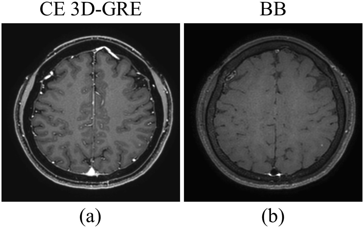
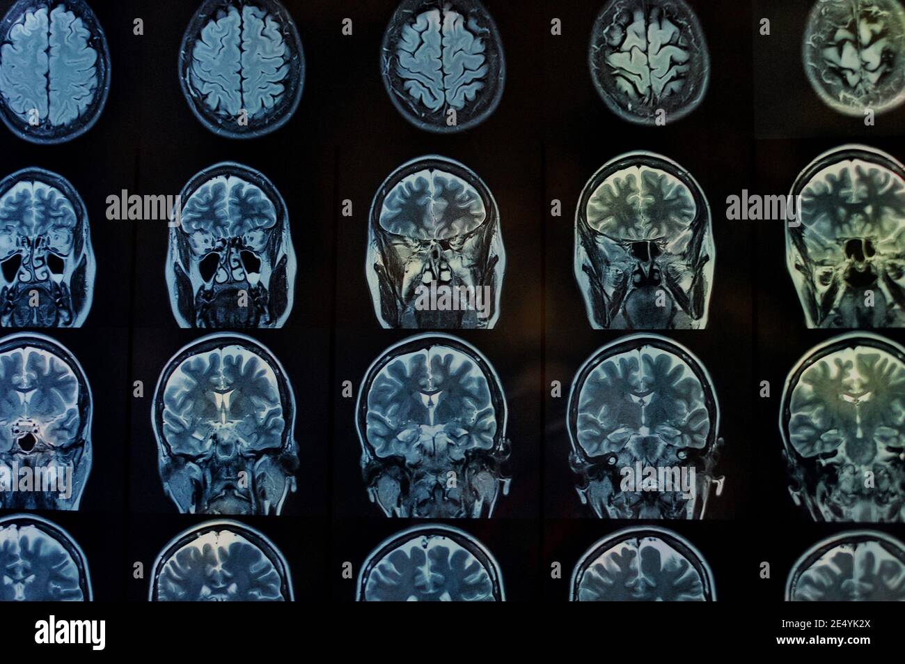
MRI brain (summary) | Radiology Reference Article - Radiopaedia MRI brain is a specialist investigation that is used for the assessment of a number of neurological conditions. It is the main method to investigate conditions such as multiple sclerosis and headaches, and used to characterize strokes and space-occupying lesions. Reference article Brain MRI: How to read MRI brain scan | Kenhub MRI is the most sensitive imaging method when it comes to examining the structure of the brain and spinal cord. It works by exciting the tissue hydrogen protons, which in turn emit electromagnetic signals back to the MRI machine. The MRI machine detects their intensity and translates it into a gray-scale MRI image. Functional MRI of the Brain > Fact Sheets > Yale Medicine The exercises increase activity in specific parts of the brain, increasing blood flow and oxygen to them. This activity lights up on the images created by the scanner, giving doctors a visible record of an exact map of the patient's brain. A normal MRI of the brain can last between 20 to 30 minutes, while the fMRI lasts between 40 to 55 minutes. 101 labeled brain images and a consistent human cortical ... - PubMed We introduce the Mindboggle-101 dataset, the largest and most complete set of free, publicly accessible, manually labeled human brain images. To manually label the macroscopic anatomy in magnetic resonance images of 101 healthy participants, we created a new cortical labeling protocol that relies on robust anatomical landmarks and minimal manual edits after initialization with automated labels.
Atlas of BRAIN MRI - W-Radiology Brain magnetic resonance imaging (MRI) is a common medical imaging method that allows clinicians to examine the brain's anatomy (1). It uses a magnetic field and radio waves to produce detailed images of the brain and the brainstem to detect various conditions (2).
Labeled imaging anatomy cases | Radiology Reference Article ... This article lists a series of labeled imaging anatomy cases by body region and modality. Brain CT head: non-contrast axial CT head: non-contrast coronal CT head: non-contrast sagittal CT head: angiogram axial CT head: angiogram coronal CT...




:watermark(/images/watermark_only_sm.png,0,0,0):watermark(/images/logo_url_sm.png,-10,-10,0):format(jpeg)/images/anatomy_term/anterior-limb-of-the-internal-capsule/Lcr25ClxiWMh7nkVlNHiwA_Anterior_limb_of_the_internal_capsule.png)










Post a Comment for "44 brain mri with labels"