43 fluorescent labels and light microscopy
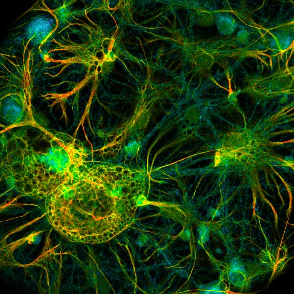
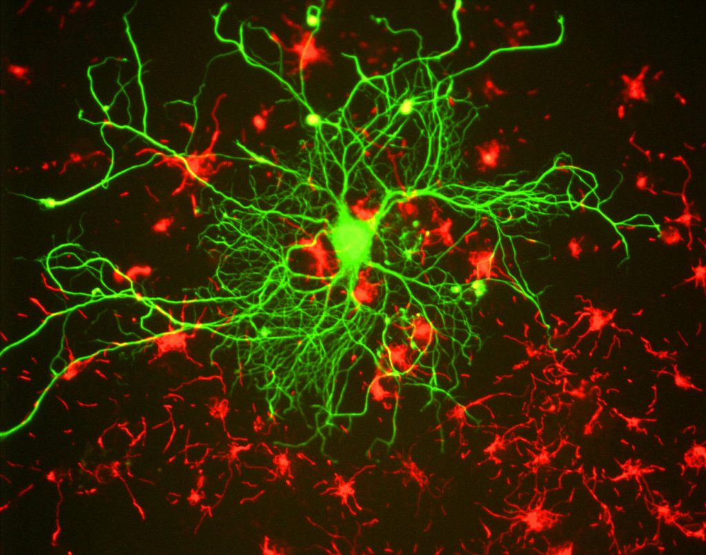
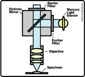
Immunofluorescence - Wikipedia Immunofluorescence is a technique used for light microscopy with a fluorescence microscope and is used primarily on microbiological samples. This technique uses the specificity of antibodies to their antigen to target fluorescent dyes to specific biomolecule targets within a cell, and therefore allows visualization of the distribution of the target molecule through the sample. Fluorescence - Wikipedia A perceptible example of fluorescence occurs when the absorbed radiation is in the ultraviolet region of the electromagnetic spectrum (invisible to the human eye), while the emitted light is in the visible region; this gives the fluorescent substance a distinct color that can only be seen when the substance has been exposed to UV light ...
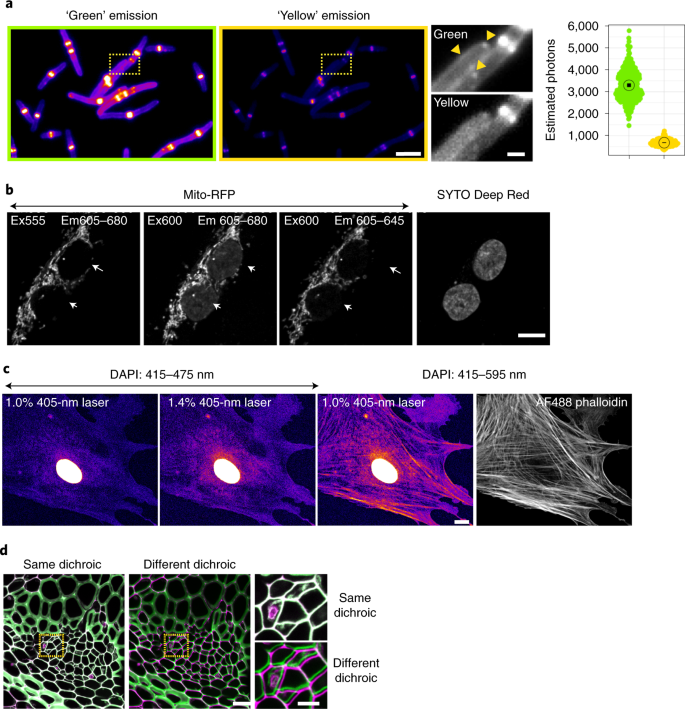
ZEISS LSM 980 with Airyscan 2 – Confocal Microscope with ... NIR fluorescent labels are less phototoxic for living samples due to the longer wavelength. This allows you to investigate living samples for longer periods of time while limiting the influence of light. Additionally, light of longer wavelength ranges is less scattered by the sample tissue, increasing penetration depth.
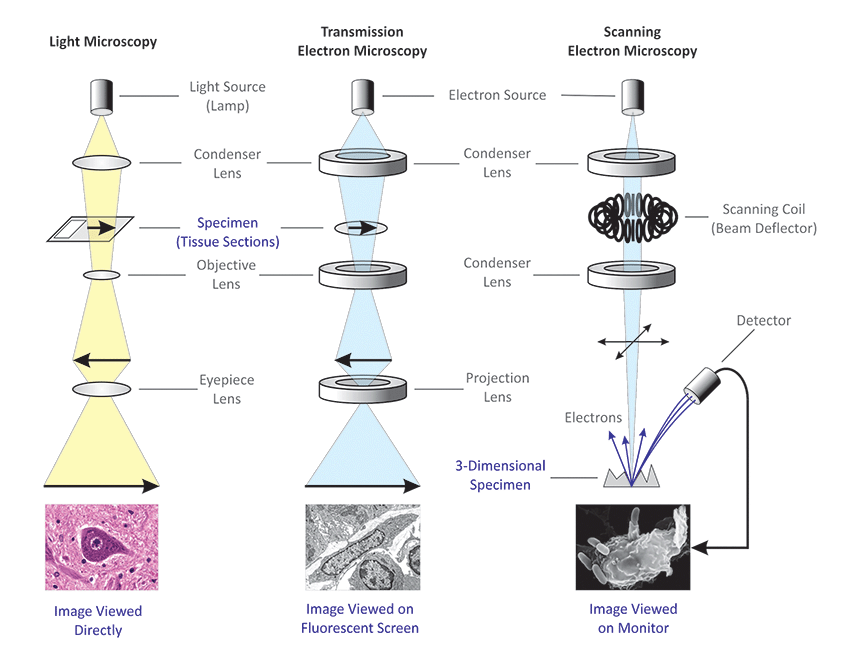
Fluorescent labels and light microscopy
Multiphoton Microscopy | Nikon’s MicroscopyU In the descanned geometry, the emitted light (illustrated in blue) returns along the same path as the excitation light, striking the scanning mirrors before passing through the confocal pinhole to the detector (Figure 4(a)). In confocal microscopy this geometry must be utilized to eliminate the detection of out-of-focus emission. Join LiveJournal Password requirements: 6 to 30 characters long; ASCII characters only (characters found on a standard US keyboard); must contain at least 4 different symbols; Two-photon excitation microscopy - Wikipedia Two-photon excitation microscopy typically uses near-infrared (NIR) excitation light which can also excite fluorescent dyes. However, for each excitation, two photons of NIR light are absorbed. Using infrared light minimizes scattering in the tissue. Due to the multiphoton absorption, the background signal is strongly suppressed.
Fluorescent labels and light microscopy. Fluorophore selection | Thermo Fisher Scientific - US Photostability in buffer is a relative measure of the percentage of initial fluorescence intensity remaining following 30 seconds of continuous illumination using a 40x/1.4NA objective and a 100-watt Hg-arc lamp as the light source with samples mounted in phosphate buffer; high photostability in buffer is ideal for live-cell imaging and detection Two-photon excitation microscopy - Wikipedia Two-photon excitation microscopy typically uses near-infrared (NIR) excitation light which can also excite fluorescent dyes. However, for each excitation, two photons of NIR light are absorbed. Using infrared light minimizes scattering in the tissue. Due to the multiphoton absorption, the background signal is strongly suppressed. Join LiveJournal Password requirements: 6 to 30 characters long; ASCII characters only (characters found on a standard US keyboard); must contain at least 4 different symbols; Multiphoton Microscopy | Nikon’s MicroscopyU In the descanned geometry, the emitted light (illustrated in blue) returns along the same path as the excitation light, striking the scanning mirrors before passing through the confocal pinhole to the detector (Figure 4(a)). In confocal microscopy this geometry must be utilized to eliminate the detection of out-of-focus emission.
















Post a Comment for "43 fluorescent labels and light microscopy"